The Therapeutic Pneumothorax
|
|
|||||
|
The Therapeutic Pneumothorax
Mario TADDEI, M.D.
FMH Specialist in Internal Medicine
Speciality: Pulmonology
Chemin des Chenevières 34
CH -2533 Evilard (Canton Berne)
Introduction
Until the discovery of antituberculous drugs (Streptomycin, WAKSMANN 1944, PAS = 4-Aminosalicylic, LEHMANN 1946 and Isoniazid, PROWAZEK and SELIKOFF 1952) tuberculosis therapy consisted of strict bed rest in sanatoriums, accompanied by a therapeutic pneumothorax (PNX) in cases of cavitary tuberculosis.
Carlo Forlanini had observed that patients whose cavities spontaneously ruptured into the pleural space sometimes showed a paradoxically beneficial course, even to the closure of the cavity.
His idea, which was revolutionary in those days (1882), was to create an artificial pneumothorax in order to immobilize the affected lung thereby hoping to achieve the same result, namely the reversal to a beneficial course. Incidentally, 1882 was the year in which Robert Koch discovered the causative organism of tuberculosis, which was later called Mycrobacterium tuberculosis. Initially it was believed that the pneumothorax immobilized the lung like a cast a broken limb. From the beginning of 1912 (Ascoli) the effectiveness of the treatment was attributed to the retraction forces of the lung.
A therapeutic pneumothorax could be induced uni- or bilaterally. In the beginning this form of therapy was restricted to unilateral therapeutic pneumothorax. It was indicated in cavitary and bacillary (open) lesions or sometimes after puncture in exudative pleurisy
Technique
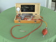
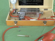
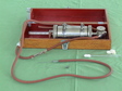
By means of devices which were improved over the course of time a certain amount of gas was introduced into the pleural cavity (MORELLI, CARPI, KüSS modified according to LESQUIN, STEINER) whereby in the beginning slowly resorbed nitrogen was used and later filtered air (see pictures). To protect the lung from injury the KüSS trocar was employed for the first insufflation. This instrument had two mandrins, a sharp point to penetrate the skin and a blunt one to perforate the parietal pleura and penetrate the pleural cavity.
The patient lay on his healthy side with a wedge-shaped bolster under the bony thorax, his arm raised and his hand under his head in order to allow access to the axial region for the procedure. The 4th, 5th or 6th intercostal cavity was punctured at the linea axillaris media. An abrupt motion of the nanometer indicator showed that the instrument had reached the pleural cavity. As a rule the pressure in the inspirium was negative (approx. 6 cm water) and in the exspirium around 0 cm.
Depending on which device was used, for example the Morelli, approx. 2/3 of 500 ml of air in a rubber balloon was aspirated, that is approx. 350-400 ml. With a piston pump 300-400 ml was also insufflated. The second insufflation was performed on the next day or the day after. As a rule the intervals were prolonged (e.g. 3, 6, 10, 15 days) whereby during the first 3 months weekly and then fortnightly on average. Only the amount was insufflated each time which was sufficient to maintain a pressure of 0 cm in the exspirium and a negative value in the inspirium. These pressure values ensured optimal expansion and ventilation of the lungs. The situation before and after insufflation was carefully monitored weekly under a fluoroscope, needless to say without an image intensifier. In retrospect the radiation dose patients were subjected to was absolutely amazing. As far as I know the total radiation dose which those patients were exposed to has never been scientifically ascertained. At the 20th International Tuberculosis Conference (Halifax, Nova Scotia) MYRDEN reported that 7.3% of the women with therapeutical pneuomothorax developed breast cancer as compared with 0.83% of a control group.
After every insufflation both interpleural pressure before and after insufflation and the amount of gas insufflated were recorded, on one hand in patient records and on the other in the patients own booklet. At greater intervals a sketch of the radiological findings was added.
Example of a Protocol:
| vor | nach | |
|---|---|---|
| 13.02.1951 PNX links | -10 | -4 (= inspir.) |
| 350 cc | ||
| -5 | 0 (= expir.) | |
| 03.03.1951 PNX links | -12 | -4 |
| 400 cc | ||
| -5 | 0 | |
| 20.03.1951 PNX links | -10 | -4 |
| 400 cc | ||
| -5 | 0 |
If the therapeutic pneumothorax could only be applied partially due to adhesions, an endoscopic adhesiolysis was performed (pleuroscopy after JAKOBAEUS).
The therapeutic pneumothorax, whether unilaterally or bilaterally, was refilled for 3-5 years. One rule stated that pneumothorax therapy was discontinued when it had been effective after 3-4 years. Effective meant cavity closure or sputum-negative.
Complications
Complications during puncture:
- Injury of the intercostal vascular or nerve bundles caused pain, minor bleeding at the puncture site, thoracic wall hematoma. Minor complications as a rule.
- Injury of the lung: frequently without recognisable consequences or limited to blood in the sputum. In rare cases tension pneumothorax.
- Vagal reaction.
- Air embolism with central nervous system symptoms. Generally avoidable if insufflation was performed when intrapleural pressure was negative.
- Subcutaneous emphysema
Pronounced dyspnoea
- Frequently caused by mediastinal hernia. Can be avoided by reducing the insufflated gas amount.
Tachycardia, palpitations are observed before action is taken.
Other complications
- Pleuritis
- Empyema
Undesirable long-term effects were also pleural synechia, pachypleuritis and pulmonary fibrosis and restrictive dysfunctions, the latter especially with bilateral therapeutic pneumothorax. Where therapeutic pneumothorax was not possible due to adhesions in the pleural cavity, occasionally a pneumoperitoneum was inserted to restrict the movement of the diaphragm thereby immobilising the lung.
Summary
Therapeutic pneumothorax was the main activity of the phthisiologist in an area when people were still dying of tuberculosis and the expression pulmonologist was not known. Later surgical procedures became successful in the treatment of TB: thoracoplasty, extrapleural pneumothorax, extrafascial apicolysis with or without plombage, oleothorax, phrenicolysis, intracavital drainage, pneumonectomy and lobectomy.
Antituberculous drugs finally rendered TB curable thus making the disease less frightening.
Besides lobectomy and segmentectomy these treatments have been abandoned and have become medical history.
Literature:
Bariéty M. and Brouet G.: Phtisiologie du médecin praticien. Masson, Paris, 1947
Bastai P. and Scarpa A.: Clinica Tisiologica. Lego, Vicenza, 1950
Omodei-Zorini A.: Corso di Tisiologia. V. Idelson, Napoli 1950
Myrden, Halifax, Nova Scotia. Actes de la XX Conférence internationale de la Tuberculose 1960
Larbaoui D.: Chimiothérapie antituberculeuse. Encyclopédie Médico-Chirurgicale,
Pneomologie, Paris, 60150A35, 1986
Written in spring 2006